

Learn about the latest research findings. Follow us on Twitter or subscribe to our RSS feed to receive our news releases. Contact our press office to be added to our media list or to arrange an interview.


Latest News
AI screening for opioid use disorder associated with fewer hospital readmissions
|
NIH-supported clinical trial shows AI tool as effective as healthcare providers in generating referrals to addiction specialists
Brain structure differences are associated with early use of substances among adolescents
|
Differences appeared to exist prior to substance use, pointing to the role brain structure may play in risk
Reported use of most drugs among adolescents remained low in 2024
|
New NIH-funded data show lower use of most substances continues following the COVID-19 pandemic
NIH and FDA leaders call for innovation in development of smoking cessation treatments
|
Engagement across stakeholders is critical to accelerate smoking cessation and reduce smoking-related disease and death
Higher doses of buprenorphine may improve treatment outcomes for people with opioid use disorder
|
Suggests higher doses of buprenorphine were associated with lower rates of future emergency department care
Fewer than half of U.S. jails provide life-saving medications for opioid use disorder
|
NIH findings highlight critical gaps in treatment access in correctional facilities for people with SUDs.
Resources for the Media
Upcoming Meetings/Events
NIDA Video News
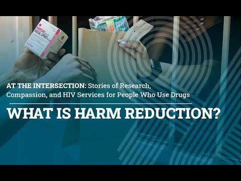

Saving Lives with Peer Support
Opioids are increasing the rates of overdose. But death is not inevitable. Peer support specialist Chetwyn “Arrow” Archer helps others connect with lifesaving care and tools, including overdose reversal medications such as naloxone.
View Transcript
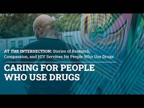
Sex, Meth and HIV
Ending the HIV epidemic requires recognizing and respecting the complexity of the health goals and lives of people who use drugs.
View Transcript
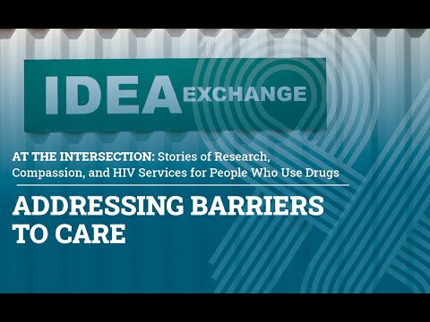
Trust, Stigma and Patient Care
Science-based care for people who use drugs includes nonjudgmental communication and goal setting. NIH-funded researchers share best practices for health professionals.
View Transcript