In this research:
- Researchers developed a tool that enables them to closely monitor HIV activity in key brain cells.
- The tool may accelerate the development of treatments for HIV in the brain.
Scientists at NIDA’s Intramural Research Program (IRP) have developed a tool to advance the study of human immunodeficiency virus (HIV) infection in the brain. It may further understanding of how HIV produces the impairments in thinking, movement, and behavior known as HIV-associated neurocognitive disorder (HAND). It also may advance the discovery and testing of treatments to alleviate HAND or to eradicate the virus from reservoirs in the central nervous system.
Dr. Lee Campbell and colleagues at IRP created the tool to enable researchers to closely monitor HIV activity in brain cells called microglia. These immune cells are the primary site of HIV infection in the brain and central nervous system. When infected by HIV, they respond by secreting inflammatory and other substances that harm neighboring neurons, causing or contributing to HAND. In addition, when HIV infects microglia, it integrates its viral DNA, or provirus, into the cells’ genome. The provirus may remain inactive or it may actively replicate, potentially releasing new viral particles into the surrounding brain tissue, causing additional cells to become infected.
The new tool couples provirus activity in microglia to production of a luminescent protein (NanoLuciferase), so that the two rise and fall together. Researchers can measure the amount of luminescent protein in the microglia and use it to compute the level of provirus activity. They can expose the microglia experimentally to different substances and conditions in vitro and track any resulting changes in proviral activity and cell function.
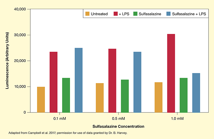
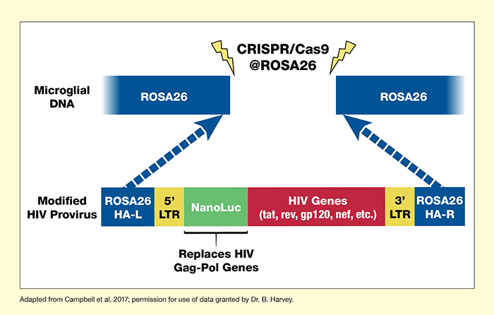
To create their tool, the IRP researchers used the CRISPR/Cas9 gene editing system to insert the HIV provirus containing NanoLuciferase into the genome of a laboratory microglial cell line (CHME5) (see Figure 1). To test the modified provirus in the microglial cells, they exposed the cells to substances known to increase or decrease proviral activity. The modified provirus responded to the exposures like normal HIV provirus, indicating that it can be used to elucidate the dynamics of normal provirus (Figure 2). Moreover, the researchers were able to manipulate and observe proviral activity with ease.
“Our research has the potential to help us both understand and treat HIV infections in the brain,” says Dr. Brandon Harvey of the IRP team. “Our long-term goals are to determine how drugs of abuse influence HIV biology in the brain and learn how to therapeutically target HIV that takes up residency in the brain.” Among research objectives that the tool may facilitate is developing therapies that permanently suppress proviral activity in microglia, a potential major step in curing the disease. Alternatively, researchers might use the tool to find ways to “wake up” inactive HIV so that it can be targeted by antiretroviral therapy (the so-called “kick and kill” approach).
This study was supported by NIH grant DA000586-05.
- Text Description of Figure 1
-
The figure shows the strategy used to create microglia containing a modified HIV provirus. The two blue rectangles at the top represent microglial DNA that has been cut apart at a site called ROSA26. The rectangle at the bottom represents the modified HIV proviral DNA. The two blue sections represent newly added DNA matching specific sections of the ROSA26 microglial DNA (ROSA26 HA-L on the left and ROSA26 HA-R on the right). The two yellow sections represent two HIV regulatory sections, 5’ LTR on the left and 3’ LTR on the right. The green section represents the newly inserted NanoLuc gene, which replaces the HIV gag-pol genes. The red section represents remaining unchanged HIV genes, including tat, rev, gp120, nef, and others. The two blue arrows indicate that the modified HIV provirus is inserted into the microglial DNA at the ROSA26 site. This is achieved using the CRISPR/Cas9 gene editing technique, indicated by the two yellow flashes.
- Text Description of Figure 2
-
This bar chart shows how the modified HIV provirus integrated into a microglial cell line responds to different activators and inhibitors. Experiments were conducted with three different concentrations of the inhibitor sulfasalazine, indicated on the horizontal x-axis. For the four bars on the left, sulfasalazine concentration was 0.1 mM, for the four bars in the middle it was 0.5 mM, and for the four bars on the right it was 1.0 mM. The vertical y-axis indicates luminescence in arbitrary units from 0 to 40,000, which serves as a measure of proviral activity. Yellow bars show activity of untreated cells, red bars show activity of cells treated with the pro-inflammatory molecule lipopolysaccharide (LPS), green bars show activity of cells treated with sulfasalazine at the concentration indicated, and blue bars show activity of cells treated with LPS plus sulfasalazine at the concentration indicated. In the group of bars on the left, untreated cells had a luminescence of about 10,000 units, cells treated with LPS had a luminescence of about 23,000 units, cells treated with 0.1 mM sulfasalazine had a luminescence of about 13,000 units, and cells treated with LPS plus 0.1 mM sulfasalazine had a luminescence of about 25,000 units. In the group of bars in the middle, untreated cells had a luminescence of 11,000 units, cells treated with LPS had a luminescence of about 25,000 units, cells treated with 0.5 mM sulfasalazine had a luminescence of about 12,000 units, and cells treated with LPS plus 0.5 mM sulfasalazine had a luminescence of about 24,000 units. In the group of bars at the right, untreated cells had a luminescence of about 11,000 units, cells treated with LPS had a luminescence of about 30,000 units, cells treated with 1.0 mM sulfasalazine had a luminescence of about 13,000 units, and cells treated with LPS plus 1.0 mM sulfasalazine had a luminescence of about 15,000 units.
Source:
- Campbell, L.A., Richie, C.T., Zhang, Y., et al. In vitro modeling of HIV proviral activity in microglia. FEBS Journal. 2017;284(23):4096-4114. DOI:10.1111/febs.14293.